
If pain is mainly on the inside of your knee and your legs are bowlegged, bone realignment may be one treatment option. In high tibial osteotomy, or HTO, the surgeon changes the path of weight through the knee to reduce pressure on the damaged compartment. Selection depends on where the wear is, the shape of the bone and your activity goals. This guide explains how HTO differs from non-surgical treatment and knee replacement, and helps you prepare for recovery.
High tibial osteotomy: who may benefit?
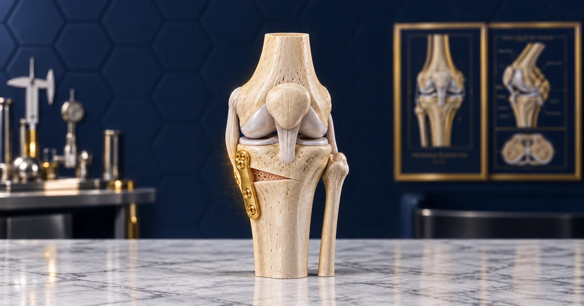
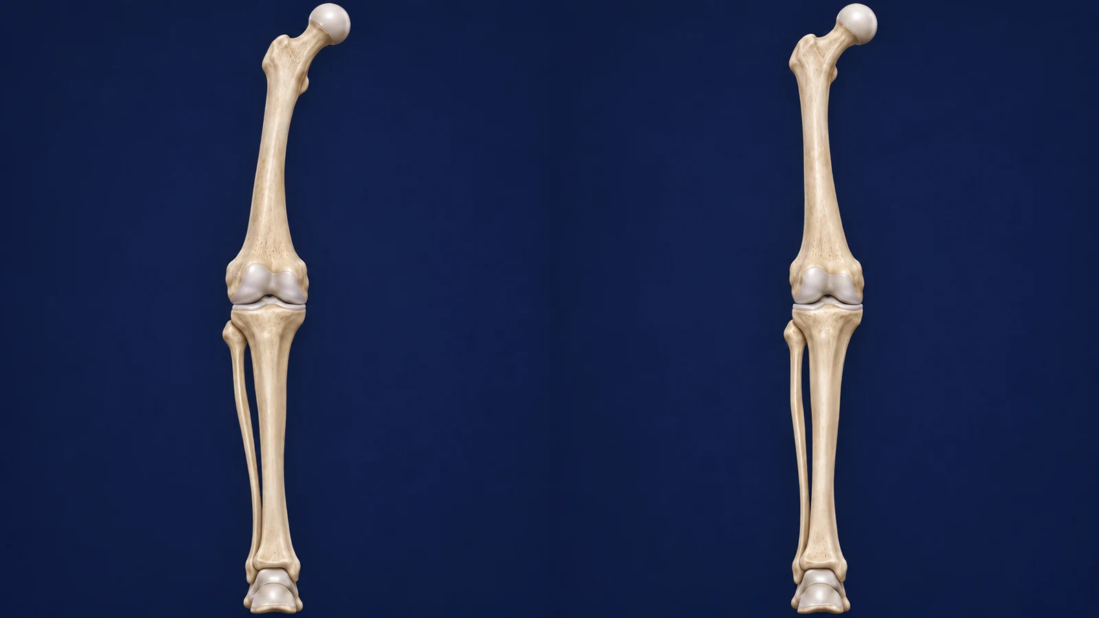
HTO changes the alignment of the upper shinbone, just below the knee. In a bowlegged knee, more load may pass through the inner compartment. If that compartment is painful and worn, a carefully planned change in bone alignment can shift load to a healthier area. The natural joint is preserved, but this operation does not automatically restore worn cartilage to its original condition.
Someone whose inner knee pain limits walking or standing, despite appropriate non-surgical treatment, may be a candidate. Knee movement, ligament health and the condition of the outer and kneecap compartments also affect selection. The appearance of bowlegs alone is not a reason for surgery; the source of pain and the likely benefit of realignment need to be clear.
Good-quality standing images of leg alignment and the knee are needed for planning. The surgeon checks whether the deformity comes from the shinbone, the thighbone or both; not every bowlegged leg should therefore be corrected by cutting the shinbone. Age or body weight alone is not an absolute reason to exclude this operation, and joint condition, general health and activity goals should be considered together.
Non-surgical treatment has a real role: tailored therapeutic exercise, weight loss when appropriate if you are overweight, activity changes and suitable medicines when needed can reduce pain and improve knee function. A walking aid or an unloading brace may also help some patients. These treatments usually do not change bone shape, but if daily life becomes manageable, a remaining deformity alone does not mean treatment has failed.
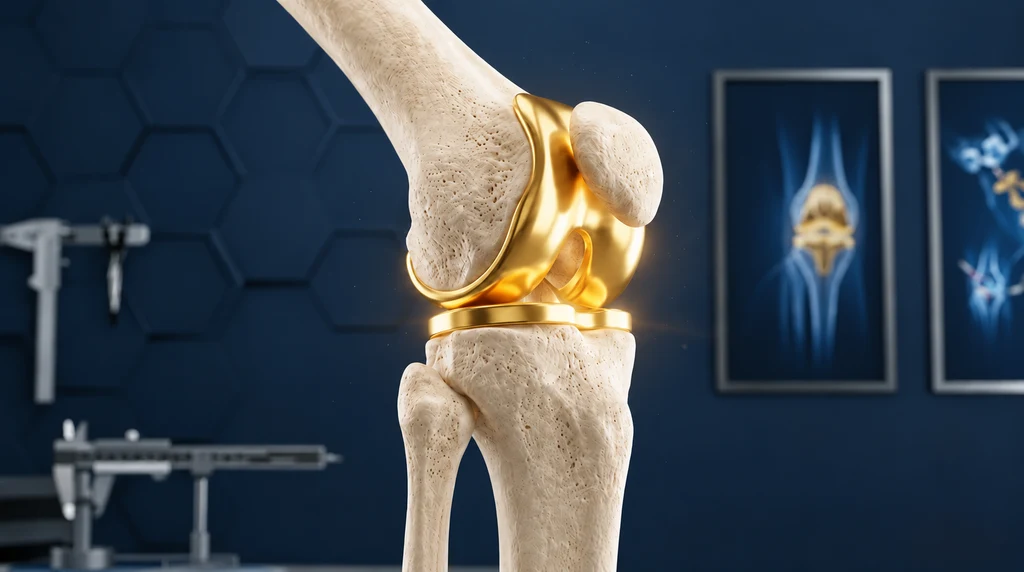
Partial knee replacement replaces the damaged surface in the affected compartment with an artificial component; HTO aims to redirect load and preserve the joint. Both are options for some people with inner-compartment osteoarthritis. Comparative studies have generally reported better pain relief and fewer complications with partial replacement, and some better movement-related outcomes with HTO, but differences between patients and techniques prevent a single recommendation for everyone. When wear is more widespread, total knee replacement should also be considered.
What if you do not have surgery yet?
In this setting, HTO is usually a planned, non-emergency operation; bowlegged alignment on an X-ray does not mean immediate surgery is necessary. If pain is manageable, you can continue non-surgical treatment and follow-up. A better decision weighs your current function, everyday priorities and the time needed for recovery. Fear of a future knee replacement alone is not a sufficient reason to rush into this operation.
In some patients, continued overloading of the inner compartment is associated with increasing pain, reduced walking ability and progression of wear. Progression is not the same for everyone, and neither leg appearance nor a single X-ray can predict when symptoms will worsen. Pain relief with non-surgical treatment also does not mean the bone has straightened or all joint wear has disappeared.
If wear becomes more extensive or knee stiffness increases, the future choice of operation may change and knee replacement may become more suitable. This possibility does not mean every delay inevitably closes the opportunity for HTO. If pain increases, your ability to work declines or you can walk a shorter distance, arrange reassessment and explain the changes in symptoms and treatments you have tried.
How can you prepare for the decision and surgery?
Before making the final decision, bring previous images and a list of treatments you have tried. Identify the activity that matters most to you: comfortable walking, standing work or sport. Ask about possible persistent pain, time using crutches and other options. You need to know whether HTO alone is planned or whether cartilage, meniscus or ligament repair is also involved; an additional procedure can change later restrictions.
If you smoke, seek help to stop before surgery; smoking can interfere with bone healing. Discuss conditions such as diabetes and heart problems during the preoperative assessment. Prepare a list of all medicines, supplements and allergies so the team can plan their use. Do not stop blood-thinning, diabetes or weight-loss medicines on your own.
At home, clear loose rugs and cables from walking routes and arrange safe access to washing and toilet facilities. Organise help with shopping, household tasks and transport. If you have stairs, practise using the prescribed walking aid safely before discharge. Tailored exercise helps osteoarthritis, but there is no single exercise prescription or repetition count for every HTO candidate; continue a programme suited to your pain and examination findings.
The day before surgery and the day itself
The day before, recheck the admission time and place, the hospital contact number and arrangements for your companion. Obtain exact fasting instructions from the anaesthetic team; permitted timing for food, water and medicines may differ between people. If medicine instructions are unclear, ask the surgical or anaesthetic team that day. Do not copy another patient's food or medicine stopping times.
For skin cleaning, follow the hospital's washing and cleanser instructions. Report a fever, a new illness or a wound or skin problem near the operation site before attending. Bring your documents, images, medicine list and comfortable clothing, and remember any prescribed walking aid. Do not drive yourself home, and arrange help in advance.
After admission, your identity and the side to be operated on are checked. The surgeon explains the correction plan and possible additional procedures, and you can ask questions before informed consent. The anaesthetic team reviews your conditions, previous anaesthetic experience and medicines. General anaesthesia and spinal anaesthesia are possible options, and the appropriate choice depends on your circumstances and the team's assessment.
The operation itself usually takes about 1 to 2 hours; preparation, anaesthesia and recovery-room time are additional. Afterwards, staff assess alertness, breathing, circulation in the foot and pain control. A longer wait for your companion does not always mean a problem has occurred; use the hospital's stated route for updates.
How is HTO performed?
Using the images, the surgeon first determines where and how much to correct the bone. The aim is not simply to bring the knees closer together in appearance; the weight-bearing path and joint surface position must also remain appropriate. If the main deformity comes from the thighbone, shinbone surgery alone may not be the right choice. This preoperative assessment helps prevent correction at the wrong site.
During surgery, a controlled cut is made in the upper shinbone and its alignment is changed according to the plan. This may involve opening a gap on one side or removing a triangular piece of bone (a wedge) from the other. The bone is then held in its new position while it heals. A special metal bone plate and screws are common fixation devices; the technique and device appropriate for you should be explained before surgery.
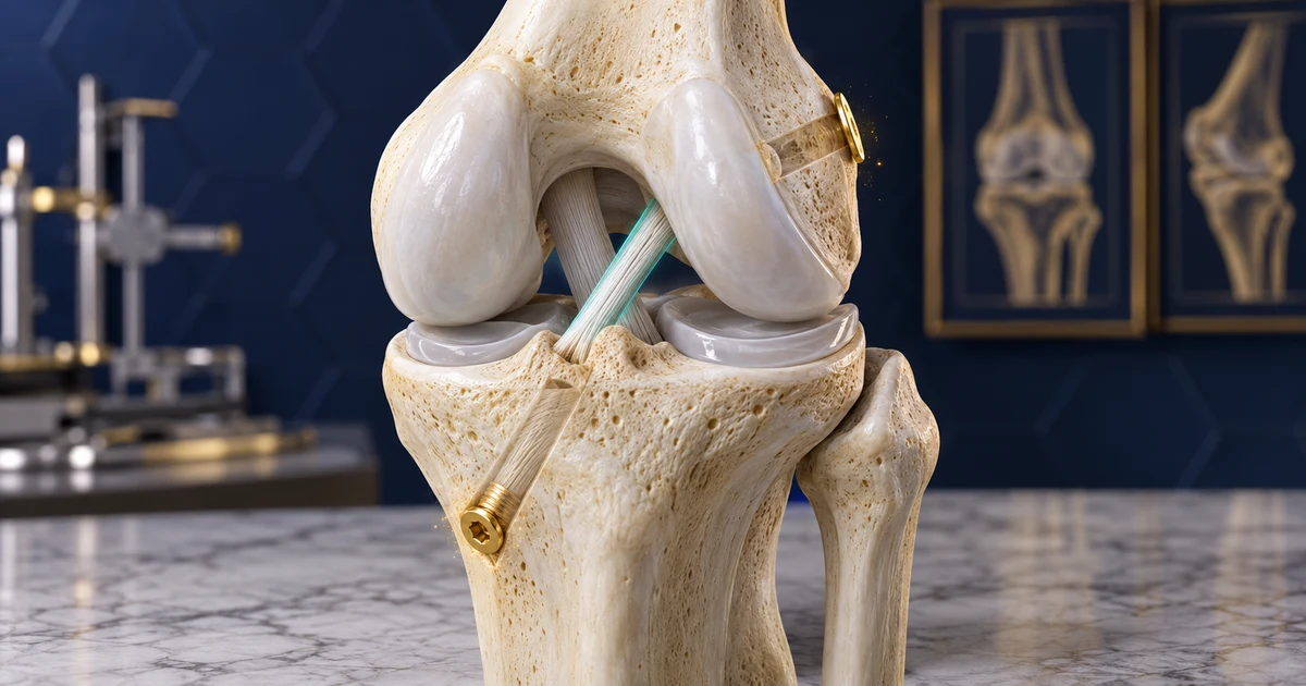
In some techniques, the gap is filled with bone graft or a substitute, but this is not necessary for everyone. If another repair inside the knee is planned, the surgeon explains it separately; not every HTO needs keyhole surgery inside the joint, called arthroscopy, or ligament repair.
As with any operation, risks include infection, blood clots, knee stiffness, nerve or vessel injury, and delayed or failed bone healing. Another operation is sometimes needed for healing problems or irritation from the fixation device. Ask about risks related to your health, smoking and type of work. Understanding these possibilities supports an informed decision and does not mean they happen to every patient.
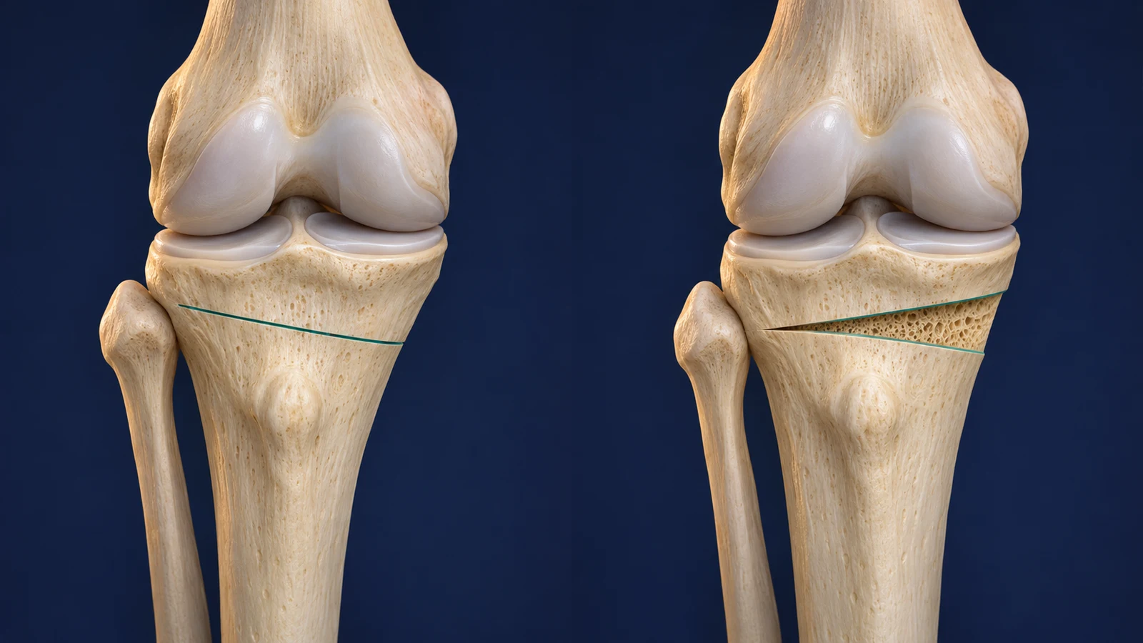
Recovery at a glance
Many patients go home about 1 to 2 days after surgery; pain control and safe mobility affect the discharge decision. Crutches and sometimes a brace are needed to protect the bone. Under the plan specified by your surgeon, use two crutches for about 6 weeks and let the operated foot make only light contact with the ground. After that, weight bearing is increased following examination and follow-up X-rays; less pain is not permission to put more weight through the leg earlier.
Returning to full activity may take about 3 to 6 months, but reaching that point on the calendar alone does not permit strenuous sport. Follow-up X-rays, bone healing, muscle strength and walking quality affect progression. Read the guide to care and rehabilitation after high tibial osteotomy for dressing, exercise and rehabilitation stages; this section is only an overview.
Contact your treatment team the same day for uncontrolled pain, persistent wound drainage or spreading wound redness. Breathlessness or chest pain, a cold foot, or severe and increasing calf pain, even without unusual tightness, need emergency assessment; do not wait for the next appointment.
Returning to everyday life: answers to 20 questions
1) When can I put weight on the operated leg?
Under the plan specified by your surgeon, use two crutches for about 6 weeks and let the operated foot make only light contact with the ground. After that, increasing weight bearing requires examination and follow-up X-rays. Less pain or simply reaching week 6 does not by itself permit putting more weight through the leg.
2) When can I stop using crutches?
Once sufficient weight bearing is allowed and you can walk without limping or losing balance, you can gradually reduce crutch use. A follow-up X-ray and gait assessment are also needed; less pain alone is not enough. Stopping crutches too early can take you beyond the permitted load.
3) When can I drive?
For planning, around 6 weeks away from driving is common in HTO guides, but that date is not automatic clearance. You must have stopped using crutches, be free of sedating medicine effects on driving, have full leg control and the ability to make an emergency stop, and sit comfortably behind the wheel. The operated side and whether the car is manual or automatic affect the decision.
4) When can I return to desk work?
For some sedentary jobs, a hospital guide suggests about 2 to 3 weeks off; some people need longer. Returning is more practical when travel is safe, pain is controlled and you can change position and elevate the leg. If prolonged sitting increases swelling, arrange remote work or a phased return.
5) How much time should I allow for standing or heavy manual work?
Full return to heavy manual work may take about 4 to 6 months. Prolonged standing, lifting, kneeling and ladder work have different demands. Start with lighter duties where possible, and assess task performance and bone healing before full duties.
6) What if I have stairs at home?
Stairs are not an absolute barrier to going home, but you should practise using them safely with your walking aid before discharge. Do not attempt stairs alone until you have adequate balance, a handrail and help. If you cannot manage them without exceeding your weight-bearing limit, temporarily arrange your resting area on one floor.
7) How should I sleep to keep the knee in a suitable position?
Sleep in a comfortable position that does not twist the knee and respects brace or movement restrictions. Support the calf and heel when resting, and do not leave a pillow only behind a bent knee for long periods. If a night brace has been prescribed, do not remove it just for sleeping comfort.
8) When can I shower?
While the dressing must stay dry, wash without wetting it. Showering is safer when you can protect the wound and sit or stand without a fall risk. Baths, pools and soaking require a closed wound and specific permission; a dressing that appears waterproof is not enough on its own.
9) When are stitches checked or removed?
The wound is usually reviewed about 2 weeks after surgery. If you have non-absorbable stitches or clips, removal depends on wound closure and the treating clinician's decision; absorbable stitches may not need removal. Before discharge, confirm the wound-review appointment and dressing type.
10) When can I fly?
One hospital HTO guide recommends waiting at least 6 weeks before flying. This does not guarantee safety for everyone; journey length, a history of clots and your mobility need assessment. Before buying a ticket, discuss fitness to travel and clot-prevention needs with the team, and check the airline's requirements.
11) When does physiotherapy start?
Safe mobility teaching usually starts in hospital, and some guides plan physiotherapy follow-up within the first 2 weeks. Knee movement and strengthening must match any additional procedure and your restrictions. The aim is to maintain movement and restore strength without putting the bone correction at risk.
12) When can I start gentle exercise?
About 3 to 6 months is an educational estimate for overall activity recovery; activities do not all restart together. A stationary bike or low-impact activity is added when knee movement and control are appropriate and healing allows it. Swimming waits until the wound is closed and clinical permission has been given.
13) Can I run or play football again?
Returning to sport is possible for some patients, but the same previous level is not guaranteed. Bone healing, strength, balance and movement control need assessment before running and pivoting sports. Remaining osteoarthritis or an additional repair may limit sport choice; reaching a particular month alone is not a starting criterion.
14) What position is better for prayer?
While kneeling, sitting on the floor or getting up would exceed movement or weight-bearing restrictions, pray safely on a chair. Returning to the floor requires the ability to lower yourself and rise with balance and without extra pressure on the knee. There is no single date for everyone, and this advice concerns knee protection.
15) When can I resume sexual activity?
A gradual return can be discussed once pain is controlled and you can change position without unintended weight through the knee, twisting or bending beyond its permitted limit. Choose a position that supports the operated leg and keeps your partner's weight off it. There is no documented fixed time for HTO; stop if movement increases pain or swelling.
16) Are pain and swelling normal after surgery?
Some initial pain, swelling and bruising are expected and should be manageable with the treatment plan. The overall trend should be towards improvement, although swelling can temporarily increase after activity. Do not assume uncontrolled or increasing pain is normal, and follow the warning signs in “Recovery at a glance”.
17) Must the fixation device be removed later?
Removal is not compulsory for everyone. If the device irritates tissues or causes pain after the bone has healed, removal may be considered. The decision depends on the site of discomfort, images and bone condition, and requires a separate operation.
18) Is the result permanent?
HTO aims to reduce pain and preserve the joint for longer, rather than stop osteoarthritis with certainty. You may not need knee replacement for years, but some patients need it in the future. Results depend on the initial joint condition, appropriate correction, healing and later activity.
19) Should both bowlegged legs be operated on together?
Bowlegs on both sides do not mean an equal need for surgery; pain, wear and the source of deformity are assessed separately for each leg. Decisions about staged or simultaneous surgery also need to consider safe mobility and help at home. Do not infer the timing or need for surgery on the other side from its appearance.
20) Why do I still need X-rays and visits when pain has improved?
Less pain does not prove complete bone healing. Images and examination assess the correction site and guide decisions about increasing load and activity. Continue follow-up even when you feel well, so progression reflects bone condition and leg function.
Summary and next step
HTO for bowlegs is useful when the source of pain and site of deformity are correctly identified, and joint preservation fits your activity goals. Non-surgical treatment, HTO and knee replacement each have a place. Bring your images, previous-treatment list and questions, and clarify the weight-bearing plan and help needed at home before giving consent.
For further reading, see care and rehabilitation after HTO and limb deformity correction options.
To discuss whether HTO may suit your knee and activity goals, call Dr Jalil Emad’s clinic on 0913 782 5207, or book an appointment online.
Read more about limb alignment correction: Limb deformity correction
This article is educational; the appropriate decision for you is made after examination and review of your images.
Scientific references
View the scientific references
- AAOS OrthoInfo. Osteotomy of the Knee. Patient guide.
- Dawson M, et al. Osteotomy around the painful degenerative varus knee has broader indications than conventionally described but must follow a strict planning process: ESSKA Formal Consensus Part I. Knee Surg Sports Traumatol Arthrosc. 2024;32(7):1891–1901. doi:10.1002/ksa.12256. PubMed.
- ESSKA Formal Consensus Part II: surgical strategy and complication management. Knee Surg Sports Traumatol Arthrosc. 2024;32:2194–2205. doi:10.1002/ksa.12273. PubMed.
- NICE. Osteoarthritis in over 16s: diagnosis and management. NG226. Recommendations.
- Ma X, Wang Y, Fu K. High tibial osteotomy versus unicompartmental knee arthroplasty for medial compartment knee osteoarthritis: a comprehensive meta-analysis with patient-specific stratification by age, BMI, and preoperative activity levels. BMC Musculoskelet Disord. 2026;27:270. doi:10.1186/s12891-026-09590-7. PubMed.
- AAHKS. Surgical Options for Knee Arthritis. Professional association patient information.
- Royal Orthopaedic Hospital NHS. High Tibial Osteotomy and Distal Femoral Osteotomy. Patient information.
- Royal College of Anaesthetists. Preparing for surgery. Preparation checklist.
- Mater. High Tibial Osteotomy. Home preparation and physiotherapist teaching only; its technique and movement protocol are not generalized. Patient information.
- Wrightington, Wigan and Leigh NHS. High Tibial Osteotomy Surgery, version 5. Work, driving and travel guidance. Patient leaflet.
- UCSF Orthopaedic Institute. Pre and Post-Operative Instructions: HTO/DFO. Dressing, resting and contact principles only; no clinic loading schedule is inferred. Patient instructions.
- Kunze KN, et al. Return to Work and Sport After Proximal Tibial Osteotomy and the Effects of Opening Versus Closing Wedge Techniques on Adverse Outcomes: A Systematic Review and Meta-analysis. Am J Sports Med. 2020;48(9):2295–2304. Epub 2019 Nov 27. doi:10.1177/0363546519881638. PubMed.
- Ekhtiari S, et al. Return to Work and Sport Following High Tibial Osteotomy. J Bone Joint Surg Am. 2016;98:1568–1577. doi:10.2106/JBJS.16.00036. PubMed.
- NHS. Pulmonary embolism. Symptoms and urgent action.
- AAOS OrthoInfo. Compartment Syndrome. Acute symptoms and emergency assessment.
- NSW Health Agency for Clinical Innovation, Emergency Care Institute. Acute limb ischaemia. Minor revision July 2026. Pathway entry and emergency assessment.
- Ohio State University Wexner Medical Center. High Tibial/Distal Femur Osteotomy Clinical Practice Guideline. Updated April 2023. Functional readiness and return-to-sport assessment principles only; no institutional loading schedule is adopted. Institutional clinical guideline.